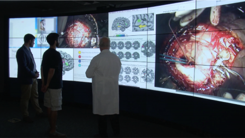
Doctors Brad Mahon and Web Pilcher look at post-surgery photographs with patient Dan Fabbio
By Dan Gross
For a brief moment, the video took over the Internet. A young man on his side with sax in hand, his brain exposed, played a beautiful melody, and the operating room cheered. But there’s more to the story. The musician, Dan Fabbio, had a tumor in his superior temporal gyrus, the area of the right temporal lobe that’s colloquially known as “the brain’s music processing center.”
I spoke with Eastman professor Betsy Marvin (Professor of Music Theory at the Eastman School of Music and Chair of the Music Theory Department) about her involvement in this incredible project, of how UR and Eastman came together not only to remove a tumor from this beloved musician and educator, but to engage in a research project that resulted in the most detailed mapping of music in the superior temporal gyrus ever done.
Betsy Marvin is a music theorist by training. She started getting interested in music cognition while working on her dissertation, which was on musical contour in 20th century atonal compositions, and how to teach students to perceive atonality; following the work of her advisor, Bob Morris, regarding musical contour (the general pitch and rhythmic shapes we perceive). Along the way, she discovered a huge amount of literature in Rush Rhees Library on contour in the perception of language. The experimental psychology apparatus was there, and it got her interested in the relationship between speech and music.
When she was a new assistant professor at Eastman, Marvin discovered the University’s “tuition benefits;” she started taking courses on River Campus. It was mostly beginning psychology courses, experimental design classes, statistics; she started to become involved in the music cognition community and running her own experiments. As she says: “That was my way in; through the back door.”
Then she had a Bridging Fellowship – only the University of Rochester offers this type of fellowship – an opportunity for faculty to take a semester off, and put themselves in residence in another field or department. Marvin took a semester off and was a student of cognitive psychology with Elissa Newport, former chair of the Brain and Cognitive Sciences Department (now at Georgetown University), and became more comfortable in the cognitive science world. When she was back at Eastman, she developed a new course (“a pretty big and popular class for mostly undergraduates,” in her words) “Music and the Mind.” Marvin has a secondary appointment in Brain and Cognitive Sciences at UR. That’s how she met Dr. Brad Mahon (Associate Professor, Brain and Cognitive Sciences, Neurosurgery, Neurology, Center for Visual Science, Center for Language Sciences and Scientific Director for Program for Translational Brain Mapping), and neurosurgeon Dr. Web Pilcher (Ernest & Thelma Del Monte Distinguished Professor of Neuromedicine and Chairman of Neurosurgery at the University of Rochester Medical Center).
How did your involvement start?
Brad phoned me up, and said, “We have a musician who’s about to undergo brain surgery, and we want to make a research project about this to assist in preserving his brain’s musical functions, and we’d like to have a music consultant.”
How long did the process take?
The surgery was on June 28, 2016, and Brad reached out to me the fall before that, in October 2015.
The superior temporal gyrus hadn’t been mapped this way before. So how did you start preparing yourself?
Musical function in the brain has been explored by a lot of other researchers before now, so I’d say there’s twenty years of good research, starting with EEGs, MEG, and fMRI… There’s a broad literature on the brain. We weren’t starting fresh. We knew that the tumor was in the right temporal lobe, and there was a danger that was in an area important for music processing. We know areas that are generally important for music, but they can differ slightly from individual to individual. The mapping that Doctors Pilcher and Mahon developed was to pinpoint those music areas in the superior temporal gyrus in Dan specifically.
Where the tumor was located, they could potentially approach from all sides, but the mapping showed that the important musical areas were in a sliver of brain located right above the tumor, so surgically they should go in from below.
So they did the tests before the surgery?
Yes. Brad got in touch with me in the fall of 2015, and the first thing we discussed was “How can we measure his musicianship before and after surgery?” I made him aware of two standardized tests of musicianship; the first is used at Eastman by faculty in our Music Teaching and Learning Department, Edwin Gordon’s Advanced Measures of Music Audiation (AMMA), and the other is the Montreal Battery for Amusia (amusia is the scientific name for tone deafness). Both tests have same format – they play you a melody and a second comparison melody, and you say whether they are the same or different. It might change in terms of rhythm or pitch. It’s testing how much you can hold in short term memory. Some of the melodies are little odd – modal instead of tonal – funny, unusual meters… I gave Brad copies of tests, and then he went away and started working with the patient, and I didn’t hear from him for many months.
In his appointments with the medical doctors, the patient discovered that the tumor was large and slow-growing, and that there wasn’t a big rush to take it out. Dr. Pilcher said he probably had had this tumor for many years, and was never aware of it until it got big enough to give him these musical hallucinations.
In working with the patient, Brad decided not to use the melodies in the format of the original tests; he thought it would be more authentic to have the patient reproduce the melodies by humming them. This is because he would also be speaking back sentences. We wanted a direct comparison of language through speech with music through humming. One of the big areas of music cognition is studying the overlap or non-overlap of music and language function in the brain; it’s sometimes called “domain specificity.” Are these tasks domain-general – do they share the same parts of the brain – or are they domain-specific?
Let’s circle back to the tests. They’re weird melodies, and it’s a tough test that challenges the brain in other ways. When you were adapting those tests, how did you figure this out?
We tried to make the task parallel to other tests they were already planning for the surgery. There are some standard awake tests that they do during this surgery, language tasks; one of them is a pictures recognition task, and there’s some reading too (for example, they show a picture of a house, with a written cue: “This is a ______”). The patient has to read them off; it’s a standard test that they use to compare this surgery with other ones. Then they had to ask, what is an analogous thing we can do for music, and be able to compare them?
Another standardized test is a repetition task; you hear a sentence, and you have to repeat it. For a parallel task we decided to listen to melody and hum it again. The test ended up being “hear the sentence, speak the sentence, hear the melody, hum the melody.” That was alternated all throughout the surgery.
One of my parts was to choose and adapt these melodies because they were instrumental tunes and not made to be hummed. There were some pianistic instrumental melodies, that were challenging because they were intended to assess musicianship. Dan and I went through these batteries of melodies, and practiced humming them, and chose which ones were more singable, and which ones were best in his vocal range, which ones were not, and so on.
Because I had to be able to assess in real time whether he got it correct or not during surgery, the melodies we chose would have to be hard enough to present a little challenge, but also singable so that I could identify an accurate performance.
I’m sure there were other pre-op tests too.
There were several steps pre-operatively; Brad Mahon and Web Pilcher had been collaborating for a while, and they developed some functional testing using pre-operative fMRIs, so that you can look at a picture of the brain and its tumor plus areas of activation for specific tasks (like music), and compare in surgery with the actual brain. They used a kind of “wand” to calibrate between the two. In the months before the surgery, there was extensive testing to see what parts of the brain he was using for language, music, motor control, for all different things, so they had these fMRI images that they could use to localize function during surgery.
They then invited me to leave the room when while they opened the scalp –
Probably for the best.
– and I had previously volunteered that this would be just fine with me. I was warned – by several people – that not only the sounds, but also the smell would be something really difficult for people observing… I hung out in the nurse’s lounge for half an hour. When they brought me in, the brain was exposed, and there was drapery all around. I have to say, the doctors and nurses were fantastic in providing an ongoing tutorial for me. They were explaining things the whole time. One thing I didn’t know was that the whole surgical arena is not sterile; there were draperies of a specific color that I couldn’t touch because there they were sterile. But the whole rest of the room wasn’t sterile; Dan had brought in his saxophone, and that was set up in the corner of the room.
I suspect this was the first time you had seen a brain in person, in a person. Was it a shock?
It was. I walked over there, and I took my first glance, and I immediately looked away and thought, “how am I going to do this?” (this was before he was awake). Then I realized in a corner of the room, they were recording it and playing video on a screen monitor for research purposes. I walked over and watched the monitor… Having the one-degree of separation looking at the monitor and not just his brain was helpful; I got accustomed to it, and then I “girded my loins,” looked at the brain, and I realized I could at that point. It was quite fascinating being that close to it.
Once the brain was exposed, and they had the wand calibrated, then before they woke him up, they tested to see how much electrical stimulation each part of the brain could withstand. They had something called a “halo,” which is a metal round ring that had electrodes dangling from it. They placed it on his brain, and they placed little numbers in various areas, so on his brain were tiny numbers that looked like they were cut out from a magazine. They systematically went through each numbered spot and stimulated it, and in real time there was an EEG so you could see the stimulation happening, and they were looking for after effects of the stimulation. And they were trying to judge what the right amount of current to send into the brain at each position.
Were they testing the whole brain, or just the superior temporal gyrus?
Just the right temporal lobe; he was lying on his left side. When they exposed the brain, it was just a circle cut from the skull to provide access to just that portion.
You called the patient’s symptoms “musical hallucinations.” Can you expand on that?
So, the thing that brought him to the doctor to start with was that he noticed that he would hear a musical tone if you slammed a door, or a linguistic syllable. Everyday sounds were perceived as something he knew wasn’t really there, and he knew that they weren’t correct.
So when Brad first called me, he talked to me about “musical hallucinations,” and when Dan described them to me, he also described linguistic hallucinations; for example a door closing would sound like a syllable. He said in the video that there were two worlds at once; he knew what the regular door-slamming should sound like, but he also heard this other thing, and it was unclear what the actual sound was… It was like parallel universes. He knew something was wrong.
One day he had a seizure at school when he was teaching, and was rushed to the hospital. All of that was the lead-up to his diagnosis. He had no idea that he had a tumor growing. He just knew that he was having these weird experiences.
For the second part of Dan’s interview with Betsy Marvin, click here.
To watch a video about the entire surgery from diagnosis to post-surgery, click here.
Betsy Marvin, along with Doctors Pilcher and Mahon, will take part in a MEL Talk called Mapping Music During Awake Brain Surgery on Saturday, October 14, from 1:30 to 3 p.m. in the UR’s Hutchison Hall, Hubbell Auditorium, River Campus. Admission is free.